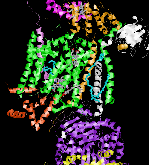

Below, on the left, is a picture of complex III in ribbon diagram. The ribbon for each polypeptide is drawn in a different color
The green part of the of the protein complex is the part that actually crosses the inner mitochondrial membrane. In fact a few hydrophobic molecules even show up in this picture.... They are the "small" molecules shown in cyan in the middle of the picture.
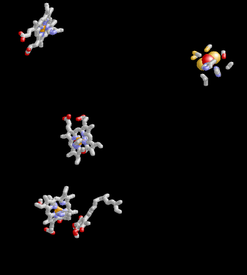
The inter membrane space would be at the top of the picture while the inner matrix would be at the bottom. Draw in stick figures are three hemes, and on Coenzyme Q molecule (all mostly on the "left side") in the upper right drawn in spheres is an iron sulfur cluster.
 |
Shown in this picture is the same orierentation of the same protein, but just the cofactors. CoQ at the bottom, 3 hemes going through the membrane region of the protein and an Fe sulfur center at the top right. |