Click an atom to diplay it's identity here
Messages about the currently highlighted features
rotate molecule (Z-axis only)
Zoom in/out
Move molecule
Java menu

 This picture shows a protein that requires 4 subunits to form a functional protein. In this case, the protein is hemoglobin (the oxygen carrying protein in blood). Each subunit is represented in the ribbon models (no amino acid atoms are shown) and colored differently. Subunit
 Some proteins require more than a single polypeptide to be a single functional unit. How these multiple polypeptides are arranged describes the
The use of the Velcro analogy again is on purpose. These polypeptides are "held" together by the same set of interactions that are important in maintaining tertiary structure.
There will be much more about how the subunits act together for form a functional unit as the course progresses. But for now I will add that they form for different reasons. Sometimes the various subunits within the protein have very different functions - each performing a part of the overall reaction (for instance). In these cases, only the total reaction of all the subunit combined make sense. we will see many examples of this throughout the metabolism portion of the course. In other cases all the subunits have the same function, but they "cooperate" in specific ways to enhance the abilities of the activity. As we will see in module 8, this most frequently occurs when the apparent rate of an enzyme must change to accomodate varying physiological states (regulation).
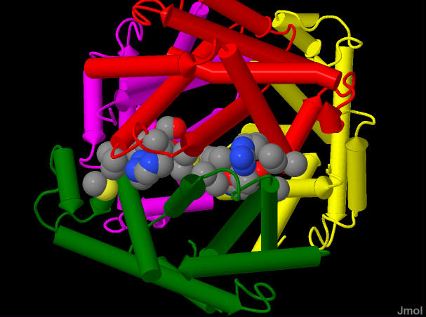 This is an image of hemoglobin showing all four subunits colored as described above, but with a few of the amino acid sidechains shaown as sheres - colored by element. The sidechains shown are those from either the
 | Hemoglobin has four subunits but only two different types. one type is called α and the other β. the common notation is that hemoglobin has an α2β2 configuration. There is a high degree of symmetry to how the protein assembles. Below three different interfaces between subunits: The | |
|
| |
|
Click an atom to diplay it's identity here | |
|
Messages about the currently highlighted features | |
|
rotate molecule rotate molecule (Z-axis only) Zoom in/out Move molecule Java menu | |