
The tertiary structure is the overall fold of the polypeptide. In other words this takes into account not only the backbone atoms and their local bend but how their local structures arrange themselves and how the sidechains of each amino acid interact. An important note here is that because the polypeptide folds you will find that some amino acids that are a long ways away from each other in sequence may or may not be near each other in SPACE. As an example in one of the blood clotting factors there are three important amino acids involved in the catalysis (MUCH more about catalysis in modules 4 and 5)
 This is the ribbon representing the mainchain atoms of factor VIIa (one of the blood clotting factors). The atoms of the sidechains are not shown so that you can see how the protein folds. notice how you can plainly see regions of α-helix, β-sheet and places of random coil. Every protein that has the same primary sequence of amino acids (there are perhaps millions of such molecules in your blood at any time) have the same fold and structure. This is a stereo image pair of Factor VIIa. If you know how to use these or have the proper "glasses" it can these images can be combined to appear to 3D.
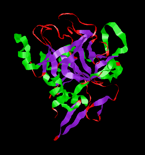
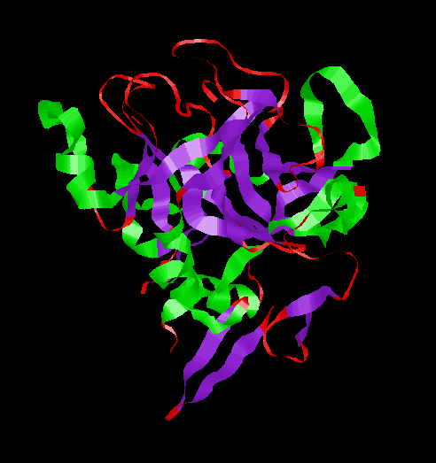
These three amino acids are actually quite far from other in the sequence but are very near each other in the folded structure.The pictures below represent the 3-D structure of Factor VIIa in a couple different ways.
only the main chain atoms are shown and they in are represented as a ribbon. The α-helices are colored green, the β-sheet regions are colored purple and the regions that are in random coil are colored red.
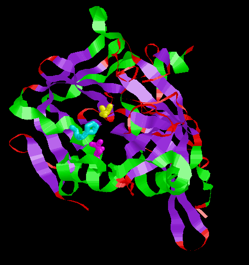 This is another view of the same factor VIIa, but now the side chain atoms of Ser195 (yellow) His57 (cyan) and Asp102 (purple) have been added. Notice the proximity of these three amino acids... even though they are not near each other in the primary sequence.
 This is another view of the same factor VIIa, but now the side chain atoms of Ser195 (yellow) His57 (cyan) and Asp102 (purple) have been added. I have animated it to rock back and forth so that you can see the 3D relationships of the various parts.
These ribbon views are perhaps a bit misleading. While they do let us see the way that the polypeptide wraps up well into a well ordered structure, it appears as if there are lots of holes in the protein. This is rarely the case. Once all the sidechains are shown in their spacefilling spheres it is clear that generally proteins "pack" their atoms rather efficiently
 Here all the (non-hydrogen) atoms of the protein are shown as spheres with diameters appropriate for their size. most atoms are colored by element type, except Ser195 (magenta) His195 (cyan) and Asp102 (purple). The yellow in this picture are sulfur atoms from cysteines. Notice that the the atoms appears to be extremely well packed together.
|
|